Cutting-edge Imaging Techniques
Digital Beam Forming (DBF)
Real-time Dynamic Aperture (RDA)
Dynamic Receiving Focus (DRF)
Tissue Harmonic Imaging (THI)
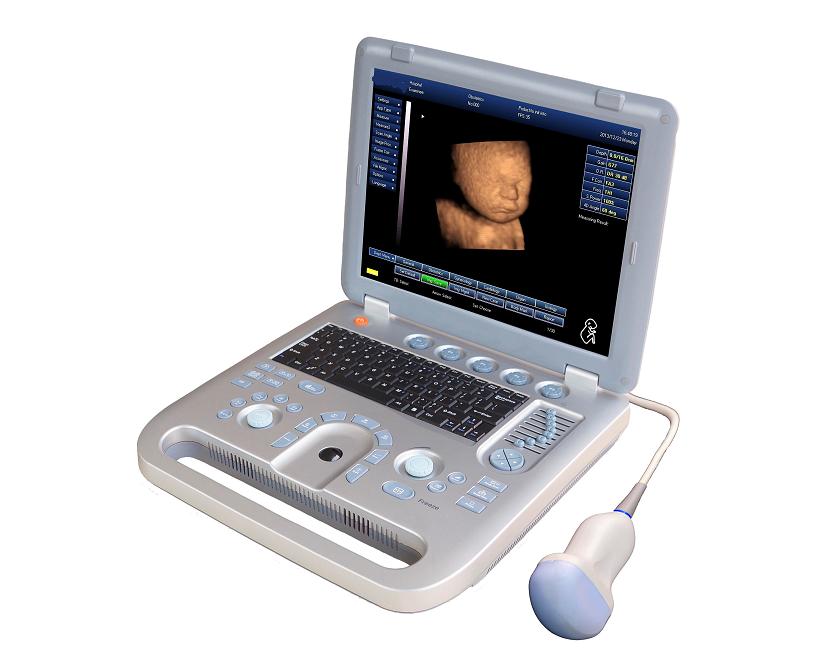
Professional Clinical Diagnostic Solutions
Applications:General, OB/GYN, Cardiology, Urology, Small organ, etc
Imaging mode:B, 2B, 4B, M, B/M
One-click optimization: Preset various calculation formula, diagnostic parameters, case library, etc
File management: Image save, video save, examination report auto-generation
Peripheral ports: USB2.0/ Video/ /S-video/RS-232/ DICOM3.0
Ergonomic Designed Keyboard, Radium Carved Soft Keys
Multi-frequency, High-density, Broadband Probes
-Volume
-Convex
-Linear
-Trans-vaginal
-Trans-rectal
-Micro-convex
Standard Configuration
-Main unit
-3.5MHz/R40 4D volume probe
-4D imaging software package
-250ml coupling gel
-AC adapter
-Built-in rechargeable lithium battery
-Protection bag
-Grounding cable
-Fuse (2)
-Operation manual
Options
-3.5MHz/R60 convex-array probe
-3.5MHz/R20 micro-convex-array probe
-7.5MHz/L40 linear-array probe
-6.5MHz/R13 transvaginal probe
-DICOM3.0
-Biopsy guide for convex probe
-Video printer
-Laser printer
-Ergonomic mobile trolley
-Air captain suitcase
-Drag and pack backbag
-Beamforming method
-DBF digital beamforming
-RDA Real-time dynamic aperture imaging
-DRA Real-time dynamic beam trace
-DRF
-DFS dynamic frequency scanning technology, 2.0-12.0 MHz operating frequency, four variable frequency scanning.
-Tissue harmonic imaging (THI)
-Display model: B、2B、4B、B/M、M
-Dynamic range: ≥100dB, 4 gears to switch
-Image processing technology: controlled frame related, gamma correction, edge enhancement, image smoothing, image noise reduction, automatic gain control, image up and down or so black and white to reversal.
-Image magnification: stepless amplification; at the same time has a real-time dynamic PIP picture in picture function of partial discharge
-Cine Loop: 512frame, Auto/manual cine loop: more screen (4b, 9b) cine loop, B/M, M mode automatically, manual cine loop
-Storage: Image storage, cine loop storage, measurement result and report can be stored, storage capacity 4G.
-Measurement and calculation: distance, ellipse circumference and area, track method to measure the perimeter and area, elliptical method of measuring surface area and volume;
-4 pieces measuring scale; ratio measurement, wire narrow ratio, surface ratio, angle measurement; automatic calculation
-Tools: puncture guiding, histogram, profile
-Menu management interface, English operating system, real-time online help and navigation prompt system, image processing and presets and optimization function.
-Comment, body mark(140 kinds for different parts option), arrow.
-Patient information management: Diagnosis case management, report print, comment library, image output(USB), import data.
-Obstetrics, gynecology, small parts, Cardiology, urology, automatic measurement software
OFD、THD、TIBIA、ULNA、AFI、LIMP、BBT、FBP
Gynaecology: uterus, endometrial thickness, ovarian volume, follicle, cervical length, diameter, cervix
Small parts: optic, thyroid, jaw and face
Cardiology: AOD, LAD, IVSTd, LVIDd, AA, LAD/AOD, LVPWd, LVIDs, EF, EF SLP, CA/CE, MVCF, CO, CI, LVMWI, AVSV, FS, (ACV), ET, SV, SI, LVMW, QMV
Urology: transition zone volume, bladder volume, RUV, prostate, kidney
-Variety of obstetric measurement reports, fetal physiology ratings and reports, fetal
growth curve.
-Gynaecology, small organs, Urology heart, urological, such as automatic report
generation system.
-The following relevant extension interface
-L VGA, S - Video, TV, Video Port
-USB2.0 Port
-RJ-45 Network Port
-Support floppy disk, hard disk, burn drive, U disk, CF card, SD card to store
-Compatible ink jet, laser, video printers, video recording.
Specification
|
MODEL |
KR-2288V |
|
Monitor |
15 inch LED |
|
Display mode |
B, B+B, B+M, M, 4B, 9B |
|
Image gray scale |
256 level |
|
Cine loop |
Auto 256 frames, Manual, 4B/9B, Auto/Manual M, B/M |
|
Image storage |
>50,000 frames |
|
Scan angle |
60-160 degrees, depending on probe |
|
Scan depth |
20mm~240mm |
|
Dynamic range |
≥100dB, 4-level adjustable |
|
Image flip |
Up/down, left/right, black/ white, 90/180/270° |
|
Zoom |
Continuously, real-time dynamic PIP |
|
Focus position |
Adjustable |
|
Focal space |
Adjustable |
|
Measurement |
Distance, circumference, area, volume, angle, heart rate, time, velocity, residual urine, etc. |
|
Software packages |
General, OB, GYN, cardiology, urinary, small organ |
|
Notation |
Date, time, name, sex, age, doctor, hospital name, full screen notation |
|
Body mark |
43 types |
|
Peripheral port |
Video, S-video, USB2.0, DICOM3.0, VGA |
|
Battery volume |
8,000mAh |
|
Power consumption (MAX) |
75VA |
|
Dimension |
35.5*36.6*7.5 CM |
|
Net Weight |
5kgs |